

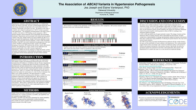
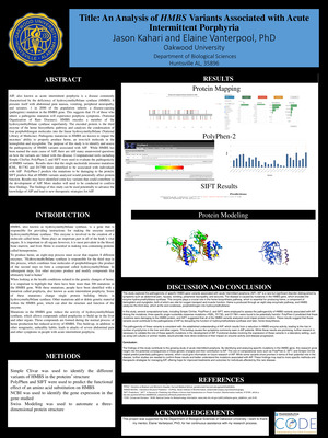
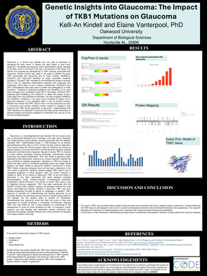

-
Effects of Cell-Free Supernatants of Commercial Yogurt Products on Salmonella Survival
Juliet Durant and Akayla King
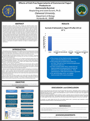
Salmonellosis remains a global public health challenge and Salmonella enterica serovar Enteritidis and Typhimurium are the primary causes. Salmonella virulence mechanisms involve attachment and invasion of intestinal epithelial cells. Gut microbiota and probiotics that contain Lactobacillus species have been shown to prevent Salmonella infection. Yogurt is a commonly consumed probiotic product containing Lactobacillus species. Yogurts made under laboratory conditions and the cell-free supernatants have been shown to inhibit Salmonella growth. The many commercial yogurt products on the market may vary in antimicrobial components based on production methods. Therefore, the present study aimed to evaluate the survival of 5 log CFU/mL of Salmonella inoculated into cell-free supernatants (CFS) of the following types of commercial yogurts: Greek whole milk plain, Greek nonfat plain, Greek nonfat vanilla, Greek zero sugar vanilla and dairy free almond milkfat. Trypticase soy broth adjusted to pH 4.5 served as the control. The CFS for all yogurt products inhibited the growth of S. typhimurium as zero CFU were observed on Tryptic Soy agar after 24 and 48 h incubation at 37°C, compared to 7.6±0.8 log CFU/mL at 24 h and with the control. Our findings suggest that commercial yogurts produce bioactive compounds that inhibit S. typhimurium growth. Because the human gut environment is complex, in vivo studies are needed to determine the impact of commercial yogurts on salmonellosis.
-
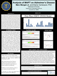
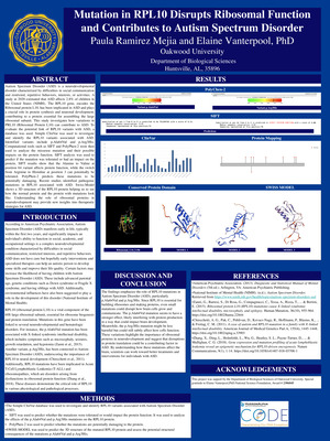
Analysis of MAPT on Alzheimer’s Disease
Elaine Vanterpool
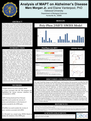
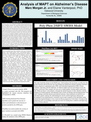
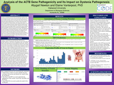
There are multiple factors that contribute to the onset of Alzheimer’s disease. These include genetic causes, environmental factors, and lifestyle choices. Microtubule-Associated Protein Tau. (MAPT) is a gene that is associated with the onset of Alzheimer’s disease. There are 2 main alleles of the APOE gene: H1 haplotype and H2 haplotype. H1 haplotype is the more common variant and is associated with an increased risk of developing tau-related neurodegenerative diseases.H2 haplotype is less common and is thought to have originated from an ancient inversion on chromosome 17q21. It may have some protective effects against certain tauopathies. The H1 haplotype is the most common, found in the majority of human populations. It is particularly prevalent in European, Asian, and African populations. Along with this the H1 haplotype has the highest risk factor for the onset of Alzheimer’s disease. If an individual has both copies of the H1 haplotype (H1/H1 genotype), they may be at a higher risk of developing tau-related neurodegenerative diseases and could experience faster disease progression compared to those who carry the protective H2 haplotype. This condition is known for progressive memory loss due to the death of neurons and a decline in brain health. This study is to gain insight into the effect that variants of the APOE gene have on the onset of Alzheimer’s disease. The MAPT gene is involved in the formation of neurofibrillary tangles (NFTs), which are a hallmark of tauopathies such as Alzheimer’s disease (AD), progressive supranuclear palsy (PSP), and corticobasal degeneration (CBD)
-
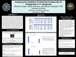
Analyzing the Inhibition Of Chemical Compounds On Collagenase in P. aeruginosa
Elaine Vanterpool
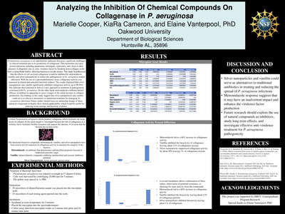
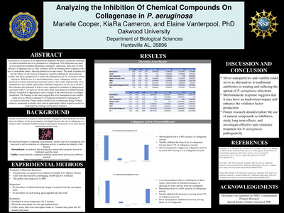
Pseudomonas aeruginosa is an opportunistic pathogen that poses significant challenges in clinical treatment due to its production of collagenase. This bacterium can cause several infections including pneumonia, meningitis, septicemia, and a host of other diseases. Collagenase acts as a key virulence factor by breaking down collagen in the host’s extracellular matrix, allowing bacteria to invade tissues. This study hypothesized that the effects of cell secreted collagenase would be inhibited by metronidazole, vanillin, and silver nanoparticles to reduce the pathogenicity of Ps. aeruginosa related infections. With the use of a spectrophotometric assay, collagenase activity was measured in treated and untreated bacterial cultures. The results displayed that silver nanoparticles and vanillin significantly inhibited collagenase activity up to 98-99%. This indicates their potential to deliver a new approach to treatment of pathogenicity associated with Ps. aeruginosa. On the other hand, metronidazole exhibited limited efficacy in addition to appearing to cause a trigger in the initial increase in collagen production. The findings in this study suggest that silver nanoparticles and vanillin could serve as effective alternatives to traditional treatment for managing Ps. aeruginosa infections. Future studies should focus on optimizing dosage of these chemical compounds to display their clinical applicability which would be useful for reducing tissue damage and bacterial virulence.
-
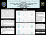
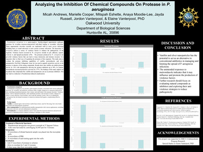
Analyzing the Inhibition Of Chemical Compounds On Protease in P. aeruginosa
Elaine Vanterpool
Pseudomonas aeruginosa is a gram-negative, bacillus-shaped opportunistic pathogen, notorious for invading immunocompromised individual leading to secondary infections. These opportunistic microbes typically are implicated mild to more severe infections including burn or wound infections to more serious systemic infections. The bacterium Ps. aeruginosa utilizes a variety of virulence factors to enhance the pathogenicity of the organism. Virulence factors secreted by Ps. aeruginosa include its pili, adhesins, capsule, exotoxin A, and lipopolysaccharide (LPS). The proteases produced by Ps. aeruginosa are powerful virulence factors that can lead to tissue destruction and immune invasion. It is imperative that we find ways of regulating the proteases of this organism.
-
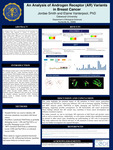
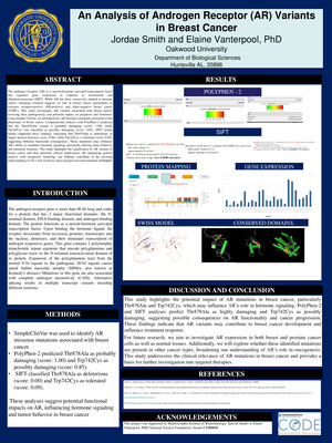
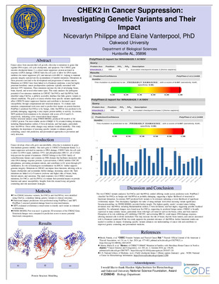
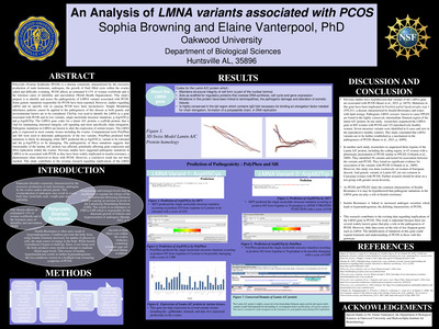
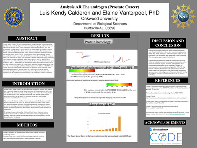
An Analysis of Androgen Receptor (AR) Variants in Breast Cancer
Elaine Vanterpool
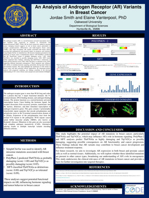
The androgen receptor gene is more than 90 kb long and codes for a protein that has 3 major functional domains: the N terminal domain, DNA-binding domain, and androgen-binding domain. The protein functions as a steroid-hormone activated transcription factor. Upon binding the hormone ligand, the receptor dissociates from accessory proteins, translocates into the nucleus, dimerizes, and then stimulates transcription of androgen responsive genes. This gene contains 2 polymorphic trinucleotide repeat segments that encode polyglutamine and polyglycine tracts in the N-terminal transactivation domain of its protein. Expansion of the polyglutamine tract from the normal 9-34 repeats to the pathogenic 38-62 repeats causes spinal bulbar muscular atrophy (SBMA, also known as Kennedy's disease). Mutations in this gene are also associated with complete androgen insensitivity (CAIS). Alternative splicing results in multiple transcript variants encoding different isoforms
-
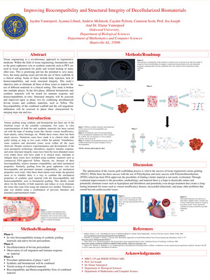
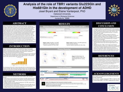
Improving Biocompatibility and Structural Integrity of Decellularized Biomaterials
Elaine Vanterpool
Tissue engineering is a revolutionary approach to regenerative medicine. Within the field of tissue engineering, biomaterials such as the great saphenous vein or synthetic materials such as PET, are used in tissue generation for grafts and wound healing in many other uses. This is promising and has the potential to save many lives, but many glaring issues prevent the use of these scaffolds in a clinical setting. Some of these include body rejection, lack of hemocompatibility, and weak structural integrity.
-
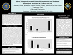
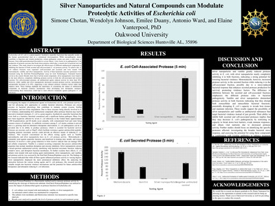
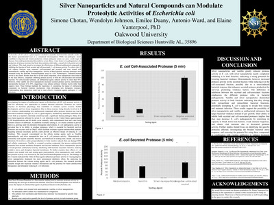
Silver Nanoparticles and Natural Compounds can Modulate Proteolytic Activities of Escherichia coli
Elaine Vanterpool
Escherichia coli (E. coli) is a gram-negative, facultative anaerobic bacterium that naturally resides in the human gastrointestinal tract as a commensal microorganism. While non-pathogenic strains contribute to digestion and vitamin production, certain pathogenic strains can cause a wide range of diseases, from mild gastrointestinal discomfort to severe illness. A key factor in its pathogenicity is the production of protease enzymes that break down proteins which play a crucial role in bacterial survival and virulence. This study aimed to investigate the effectiveness of different antibacterial compounds in inhibiting the function of both secreted and cell-associated E. coli protease activity.
-
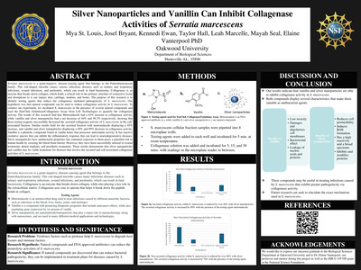
Silver Nanoparticles and Vanillin Can Inhibit Collagenase Activities of Serratia marcescens
Elaine Vanterpool
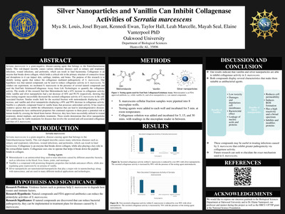
Serratia marcescens is a gram-negative, disease-causing agent that belongs to the Enterobacteriaceae family. This rod-shaped microbe causes many infectious diseases such as urinary and respiratory infections, wound infections, and peritonitis, which can result in fatal bacteremia. Protease is a virulence factor of S. marcescens that enhances its pathogenicity by breaking down proteins via cleavage of peptide bonds. The purpose of this research is to identify testing agents that reduce the protease mediated pathogenicity of S. marcescens
-
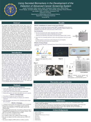
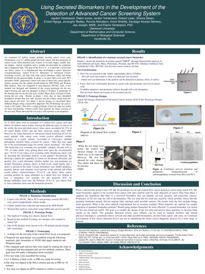
Using Secreted Biomarkers in the Development of the Detection of Advanced Cancer Screening System
Elaine Vanterpool
An estimated 20 million people globally develop cancer every year. Furthermore, over 9.7 million people die from cancer. The best prognosis of cancer occurs when detected early. Cancer, in its early stages, usually does not display clinical symptoms and is usually not detectable by symptoms and is diagnosed later. The goal of the D.A.C.S. is to provide a screen that can detect cancer. It is hypothesized that developing a cancer detection system/technology, named D.A.C.S. (Detection of Advanced Cancer Screening) system, can help with early cancer detection, while still being affordable for the general public.
-
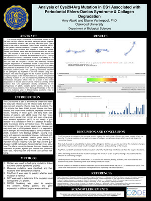
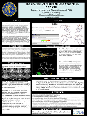
Analysis of Cys294Arg Mutation in CS1 Associated with Periodontal Ehlers-Danlos Syndrome & Collagen Degradation
Elaine Vanterpool and Amy Aboki
C1s enzyme plays a crucial role in the immune system so that the body can fight infection and repair damaged tissues. However, if it is not working properly, it will do more harm than good. This is shown in the case of periodontal Ehlers-Danlos syndrome (pEDS), a rare genetic disorder whereby C1s breaks down collagen I, a protein that gives structure and strength to connective tissues. Severe gum disease, loose teeth and premature teeth is a result of this. The purpose of this study is to identify and analyze the pathogenicity of C1s mutations and association in pEDS.
-
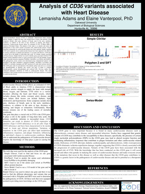
Analysis of CD36 variants associated with Heart Disease
Elaine Vanterpool and Lemanisha Adams
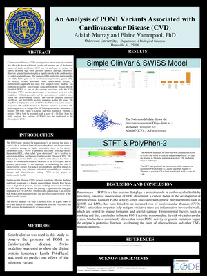
Cardiovascular Disease (CVD) affects an estimated 47-54% of Black adults in America. CVD is characterized when coronary arteries struggle to supply the heart with enough blood, oxygen, and nutrients. CVD refers to a various disorders affecting the heart and blood vessels, which includes disease like stroke, coronary artery disease, and heart failure. Risk factors can include diabetes, high cholesterol, and hypertension. Some symptoms include chest pain, shortness of breath, pain in the neck, numbness, weakness, or coldness in the arms or legs, dizziness or fainting, and fatigue or exhaustion. Unfortunately, Heart disease remains one of the leading causes of mortality worldwide. One gene that plays a crucial role in CVD is the CD36 gene. It plays a role in the uptake of long-chain fatty acids, the primary metabolic substrate in myocardial tissue. CD36 mutations can result in absence of CD36 expression in platelets (Type I CD36 deficiency) or in all cells (Type II CD36 deficiency) and have been characterized to be mostly found in Asian and African populations. Variations or mutations in the CD36 gene can affect lipid metabolism, inflammatory responses, and plaque formation, influencing an individual's chances to cardiovascular conditions. Being able to understand the pathogenesis of Cardiovascular disease requires the knowledge of what plays a part in causing the disease.
-
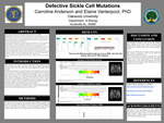
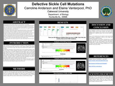
Defective Sickle Cell Mutations
Elaine Vanterpool and Carroline Anderson
Sickle cell disease (SCD) is a group of inherited red blood cell disorders. It is an inherited hemoglobinopathy. Both alleles must be affected to manifest the disease. In sickle cell disease, the red blood cells become hard, sticky, and look like a sickle, making it difficult to pass through the blood vessels and carry oxygen. Sickle cell trait is more prevalent than sickle cell disease and affects 1 in 13 African American babies (According to the CDC). The clinical manifestation of sickle cell trait is not as aggressive and does not cause as much morbidity as sickle cell disease. This study aimed to identify the gene variants associated with sickle cell disease. To do this, we used Simple Clinvar, PolyPhen-2, and SIFT to do the analysis. Simple Clinvar was used to look up sickle cell and provide information on the disease. PolyPhen-2 was used to gain visuals on the mutations, as well as information on whether or not the mutation is benign. SIFT was used to confirm whether or not the mutation is benign. The variants that were chosen were the His114Arg variant and the Glu91Lys variant. These variants were chosen at random. After some analysis from PolyPhen-2, both variants were shown to be benign. SIFT predicted that the mutations do not affect protein function. From Simple Clinvar, 423 missense mutations were identified. There are 423 mutations that can manifest in sickle cell trait. The SNV mutation is consistently in sickle cell trait. Within the SNV mutation, there are different variants. One of the drawbacks in this research is that there are so many variants. Because of this, the research is ongoing.
-
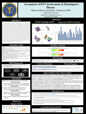
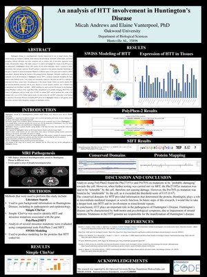
An analysis of HTT involvement in Huntington’s Disease
Elaine Vanterpool and Micah Andrews
Huntington’s disease is a neurodegenerative disorder which affects every three to seven out of 100,000 individuals. Huntington’s is categorized by behavior change, such as reward perception, motivation, decision making, self perception, and struggle with interpersonal skills. Personality change, such as isolation, anxiety, and depression are also commonly seen in cases of Huntington’s. Even though dietary restrictions and physical exercise can slow the progression of Huntingtons, stress has shown to significantly contribute to the manifestation of Huntingtons disease. •Huntington’s disease is a genetic disorder causing atrophy of the brain over time, more specifically cortical and subcortical gray matter. •Most impacted by Huntington’s disease are the striatal neurons, found in subcortical gray matter. •Impact to these striatal neurons can also cause motor issues, such as involuntary jerking, muscle contractions, unusual eye movements, and trouble with speech or walking. •The HTT gene is essential in encoding for the protein, Huntingtin. This protein is essential for chemical signaling, transporting materials, attachment to proteins and other structures, and protecting the cell from apoptosis. •Proper functionality of HTT is necessary for normal function. •In cases of Huntington’s disease, the HTT gene has been mutated. •The purpose of this study was to identify these mutations and the effects they have on the pathogenicity of Huntington’s disease.
-
An analysis of HTT involvement in Huntington’s Disease
Elaine Vanterpool and Micah Andrews
Huntington's disease is a neurodegenerative disorder characterized by loss of striatal neurons. These striatal neurons are essential in cognition, motor function, decision-making, motivation, reinforcement, and reward perception. Affected individuals may show symptoms such as isolation, lack of motivation, depression, mood swings, and personality changes. This study’s purpose is to analyze and highlight the variants of the HTT gene in the pathogenesis of Huntington’s disease.
-
Analyzing the Inhibition Of Chemical Compounds On Protease in P. aeruginosa
Elaine Vanterpool, Micah Andrews, Marielle Cooper, Mitspah Eshette, Anaya Moodie-Lee, Jayda Russell, and Jordon Vanterpool
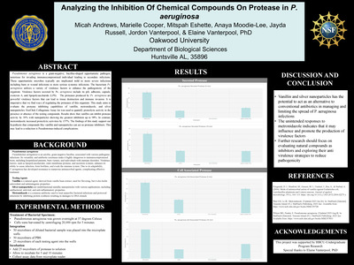
Pseudomonas aeruginosa is a gram-negative, bacillus-shaped opportunistic pathogen, notorious for invading immunocompromised individual leading to secondary infections. These opportunistic microbes typically are implicated mild to more severe infections including burn or wound infections to more serious systemic infections. The bacterium Ps. aeruginosa utilizes a variety of virulence factors to enhance the pathogenicity of the organism. Virulence factors secreted by Ps. aeruginosa include its pili, adhesins, capsule, exotoxin A, and lipopolysaccharide (LPS). The proteases produced by Ps. aeruginosa are powerful virulence factors that can lead to tissue destruction and immune invasion. It is imperative that we find ways of regulating the proteases of this organism. This study aims to evaluate the protease inhibiting capabilities of vanillin, metronidazole, and silver nanoparticles. EnzChek Collagenase Assay kit was used to quantify proteolytic activity in the presence or absence of the testing compounds. Results show that vanillin can inhibit protease activity by 30% with nanoparticles showing the greatest inhibition up to 98%. In contrast, metronidazole increased proteolytic activities by 157%. The findings of this study support our hypothesis that compounds like vanillin and nanoparticles can act as protease inhibitors. This may lead to a reduction is Pseudomonas-induced complications
-
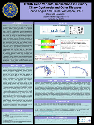
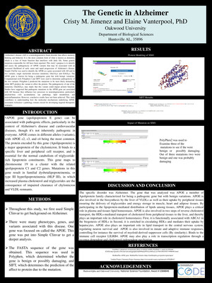
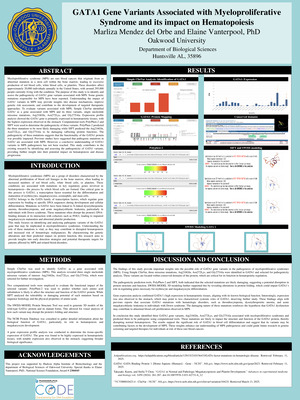
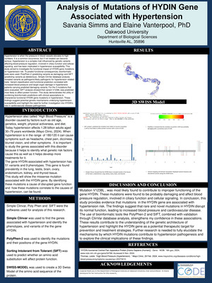
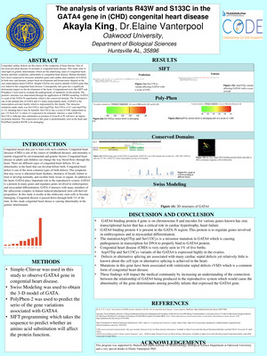
HYDIN Gene Variants: Implications in Primary Ciliary Dyskinesia and Other Diseases
Elaine Vanterpool and Sharie Angus
The HYDIN gene, also known as CILD5, HYDIN1, HYDIN2, and PPP1R31, encodes a protein that plays a role in cilia motility. Mutations in HYDIN are associated with autosomal recessive primary ciliary dyskinesia-5 (PCD5), a disorder characterized by cerebrospinal fluid accumulation in the brain’s ventricles. A duplicate copy of this gene has also been identified on chromosome 1 in humans. Research indicates that HYDIN loss of-function mutations contribute to asthenoteratozoospermia, leading to structural defects in sperm flagella, disassembly of the acrosome and neck, and subsequent male infertility. Intracytoplasmic sperm injection (ICSI) has been proposed as a potential treatment for sperm immobility caused by these mutations. Beyond its role in ciliary function, HYDIN has been implicated in other diseases. Single nucleotide polymorphisms (SNPs) in HYDIN have been linked to atrial septal defects (ASD), a common congenital heart condition. Additionally, emerging studies suggest that HYDIN variants may serve as cancer associated antigens recognized by adaptive immunity, indicating a potential role in oncogenesis. Structurally, the HYDIN protein contains conserved domains critical for cellular function. The ASH domain, present in proteins involved in cilia, flagella, the centrosome, and the Golgi complex, is also found in HYDIN and OCRL—deficiencies of which are associated with hydrocephalus and Lowe oculocerebrorenal syndrome (OCRL), respectively. This suggests that HYDIN may have functions beyond mitotic spindle regulation, potentially influencing ciliary and flagellar dynamics. Another key domain, the Trichohyalin-Plectin Homology (TPH) domain, is linked to mitochondrial movement and cytoskeletal interactions, further highlighting HYDIN’s significance in cellular processes and disease pathology.
-
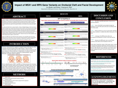
Impact of MSX1 and IRF6 Gene Variants on Orofacial Cleft and Facial Development
Elaine Vanterpool and Lisa Banks
A cleft lip or palate is a congenital anomaly that occurs when a baby's mouth doesn’t form properly during pregnancy. It happens when the tissues that shape the upper lip or roof of the mouth fail to join during development. It is among the most common birth defects associated with genetic conditions or syndromes. Orofacial cleft can be caused by a combination of genes and other factors like the mother's exposure to environmental things, diet, and medications during pregnancy. Examining the genes involved and making connections that provide insight into the improper formations of the maxillary tissues during fetal stages is crucial. This study used Simple Clinvar, Uniprot, Polyphen 2, SIFT, and SWISS Modeling to identify and analyze mutations. The genes in the orofacial cleft I chose were MSX1 and IRF6. The MSX1 gene is part of a larger family of homeobox genes, which control the formation of many body structures during early development. IRF6 gene encodes a member of the interferon regulatory transcription factor (IRF) family important for early growth. Researchers believe that deleting the MSX1 gene disrupts the formation of oral structures during fetal stages, leading to dental abnormalities. It can cause dramatic developmental defects like the loss of specific structures. The IRF6 protein is active in cells that give rise to tissues in the head and face. Computational tools PolyPhen-2 and SIFT predict MSX1 variants Ser34Arg, Gly122Glu, Ala200Val. IRF6 variants include Ala2Val, Arg84Cys, and Pro172Ser. Variants in the IRF6 gene cause popliteal pterygium syndrome. A loss of the MSX1 gene may cause an opening in the roof of the mouth, as seen in some people with Wolf-Hirschhorn syndrome. These findings could impact future research and the medical community by explaining the contributions of MSX1 & IRF6 to structural development impairments and facial defects due to pathogenic variants
-
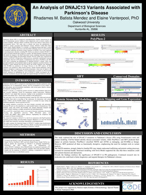
An Analysis of DNAJC13 Variants Associated with Parkinson’s Disease
Elaine Vanterpool and Rhadames M. Batista-Mendez
Parkinson's disease (PD) is a progressive neurodegenerative disorder characterized by the loss of dopaminergic neurons, leading to motor impairments such as tremors, rigidity, and postural instability. Affecting millions worldwide, PD has been linked to both genetic and environmental factors. This study aims to identify and assess the pathogenicity of DNAJC13 variants associated with Parkinson's disease.
-
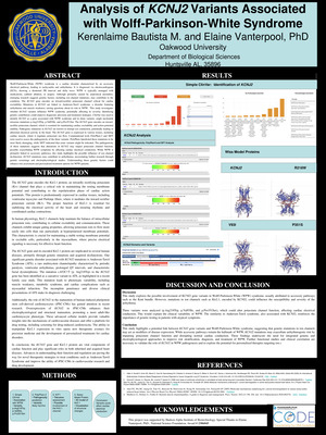
Analysis of KCNJ2 Variants Associated with Wolff-Parkinson-White Syndrome
Elaine Vanterpool and Kerenlaime Bautista
Wolff-Parkinson-White (WPW) syndrome is a cardiac disorder characterized by an accessory electrical pathway, leading to tachycardia and arrhythmias. It is diagnosed via electrocardiogram (ECG), showing a shortened PR interval and delta wave. WPW is typically managed with medications, catheter ablation, or surgery. Although primarily caused by anatomical anomalies, emerging research suggests genetic factors, including ion channel mutations, may contribute to the condition. The KCNJ2 gene encodes an inward-rectifier potassium channel critical for cardiac excitability. Mutations in KCNJ2 are linked to Andersen-Tawil syndrome, a disorder featuring arrhythmias and muscle weakness, raising questions about its role in WPW. This study investigates whether KCNJ2 variants influence WPW syndrome, potentially affecting its severity.
-
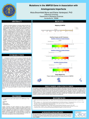
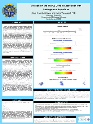
Mutations in the MMP20 Gene in Association with Amelogenesis Imperfecta
Elaine Vanterpool and Alicia Broomfield-Myrie
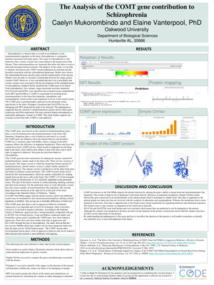
Enamel, the hardest substance in the body, protects the teeth from various forces like temperature and physical factors that would cause damage. Depending on the kind of AI one has developed, the phenotypes can range from thin teeth to teeth discoloration or decay. MMP20, a gene commonly associated with AI, encodes for Enamelysin, a protein crucial for enamel formation. It is also known as matrix metallopeptidase 20. It becomes active when it is cleaved by cellular proteases. When this gene is mutated, the splicing pattern is altered. Consequently, this can cause disease. This study was developed to analyze the pathogenicity of the mutated MMP20 variants associated with AI.
-
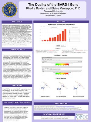
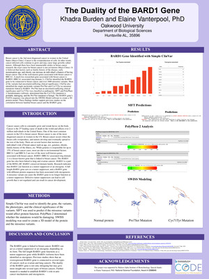
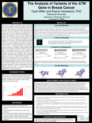
The Duality of the BARD1 Gene
Elaine Vanterpool and Khadra Burden
Breast cancer is the 2nd most diagnosed cancer in women in the United States (Mayo Clinic). Cancer is the overproduction of cells. In other words, cancer-infected cells continue to grow and may cause large growths called tumors. Although there have been innumerable strides in breast cancer research, the exact cause for breast cancer is not yet known (Mayo Clinic). It is believed that factors such as family history of the disease, early menstruation age, and obesity can increase an individual’s chances of having breast cancer. One of the well-known genes associated with breast cancer is BRCA1. A much less researched gene associated with breast cancer is BARD1 (BRCA1 associated ring domain 1). ClinVar identified the BARD1 gene to be connected to breast cancer, and over 1000 missense variants. Most of the variants had uncertain/conflicting clinical significance. ClinVar also identified the single nucleotide variants Pro7Ser and Cys71Tyr as missense mutations linked to BARD1. Pro7Ser had an uncertain/conflicting clinical significance and Cys71Tyr was classified as pathogenic. SIFT and PolyPhen2, bioinformatics software, determined that the Cys71Tyr mutation is probably damaging, and the Pro7Ser mutation is benign. The BARD1 gene’s FASTA sequence was placed into the SWISS-Model software to create a 3D protein model. These findings further support previous studies on the correlation between familial breast cancer and the BARD1 gene.
-
The Duality of the BARD1 Gene
Elaine Vanterpool and Khadra Burden
Cancer causes cells to constantly grow and wreak havoc on the body. Cancer is the 2nd leading cause of death in the world and affects over 2 million individuals in the United States. One of the most common cancers in the US is breast cancer. Breast cancer is one of the most diagnosed cancers in women in the US. Breast cancer occurs when the breast cells overproduce, and tumors develop and eventually spread to the rest of the body. There are several factors that increase an individual’s risk of breast cancer such as age, sex, genetics, obesity, family history of the illness, etc. While genetics is responsible for up to 15% of breast cancer cases, most are due to environmental factors. BRCA-1 and BRCA-2 are two of the most well-known genes associated with breast cancer. BARD1 (BRCA1 associated ring domain 1) is a lesser-known gene that is linked to breast cancer. The BARD1 gene has also been linked to lung and ovarian cancers. BARD1 is a part of the RING_HC BARD1 conserved domain family. Research indicates that BARD1 can function as a tumor suppressor or an oncogene. A full length BARD1 gene acts as a tumor suppressor, and a BARD1 gene with different protein sequences has been associated with oncogenesis. A missense variant can cause the BARD1 gene to no longer function as a tumor suppressor. Defective tumor suppressors can lead to cell growth that is not regulated and can result in cancer development.
-
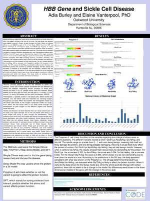
HBB Gene and Sickle Cell Disease
Elaine Vanterpool and Adia Burley
Around 100,000 people in the US are affected by sickle cell disease. There are 8 million people worldwide who are affected by Sickle cell disease. Regarding African ancestry or those who identify as black, 9 of 10 people would have this disease. Black babies can be born with this trait, and the ratio is 1 in every 13, and around 1 in every 365 babies are born with the disease. (NHLBI) •Hemoglobin, a specific protein in red blood cells, affects sickle cell disease. Our red blood cells are important for carrying oxygen throughout our body. When we breathe, the hemoglobin inside our red blood cells binds to the oxygen molecules inside our lungs. From there, the red blood cells in our body travel through our bloodstream to give oxygen to the different organs and tissues inside the body. •Sickle cell disease is a blood disorder that can cause severe pain in the body and organ damage due to the red blood cells being in a sickle shape and trying to pass through the bloodstream. Other problems that could occur from sickle cell anemia include terrible pain, blood blockages, and other health problems. Some tissues that this can affect are our adrenal tissues, heart tissue, intestines, lungs, kidneys, and stomach. It has been shown to affect the heart, lung, and stomach tissue most profoundly out of these tissues. Some complications can include certain things such as chronic pain, lung problems, as well as eye problems, infections, and even kidney diseases (NIH). It can also lead to blood cells being ruptured, anemia, immunodeficiency, and, in some cases, early death. Sickle Cell is a disease that can be very serious and affect many lives. Increasing our awareness and advancing our reach of HBB and its effect on Sickle Cell disease will be crucial for enhancing our understanding in the future
-
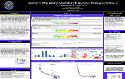
Analysis of DMD Variants Associated with Duchenne Muscular Dystrophy
Elaine Vanterpool and K’sia Byass
Duchenne Muscular Dystrophy is an X-linked recessive disorder that causes muscle weakness. This disease is characterized by frequent falls, difficulty walking, running, jumping, or scoliosis. Duchenne Muscular Dystrophy 1 in every 5000 boys worldwide causes a gene tic mutation that prevents the production of dystrophin. This study aims to identify and assess the pathogenicity of DMD variants associated with Duchenne Muscular Dystrophy. Genetic mutations responsible for Duchenne Muscular Dystrophy have been reported. Studies regarding DMD and its specific role in causing Duchenne Muscular Dystrophy have been reported to be accurate and effective. X-linked inheritance patterns are applied to this disease's pathogenesis and genetic and environmental factors. ClinVar was used to identify the DMD as a gene associated with Duchenne Muscular Dystrophy and the multiple variants, single nucleotide missense mutations, p.Lys765Glu, p.His446Arg, and p.Thr715Ser. The DMD gene codes for a dystrophin cytoskeletal protein, a scaffold protein that is a crucial link between sarcolemma and actin cytoskeleton that helps stabilize muscle fibers during contraction. Pathogenic mutations in DMD are known to alter the expression of specific muscle tissues. The DMD protein is expressed in most skeletal and heart muscles. Computational tools SIFT and PolPhen-2 were used to determine the pathogenicity of the three variants.SIFT predicted the p.Lys765Glu to be damaging, p.His446Arg to be tolerated, and p.Thr715Ser to be tolerated. On the other hand, Polphen-2 predicted p.Lys765Glu to be damaging, and the other two mutations were benign. The pathogenicity of these mutations suggests that the functionality of the dystrophin cytoskeletal protein was affected, which also potentially affects the gene expression and the X chromosome within the spine. Previous studies have connected pathogenic mutations in DMD to Duchenne Muscular Dystrophy as they have implicated in dilated cardiomyopathy, which is observed in those with Duchenne Muscular Dystrophy.
-

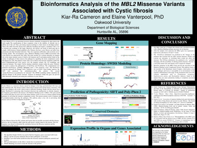
Bioinformatics Analysis of the MBL2 Missense Variants Associated with Cystic fibrosis
Elaine Vanterpool and Kiar-Ra Cameron
Cystic fibrosis (CF) is an autosomal recessive disease commonly recognized by thick mucus and loud coughs. The manifestation of these symptoms is due to the inability of chloride ions to diffuse out of the cell. Thus, preventing osmosis resulting in a thick mucus on the lung’s surface. Those who suffer from this disease have difficulty breathing and require a modulator with a vest to increase gas exchange in the lungs. Moreover, the disease can result in much pain due to coughs, which destroys ciliated epithelial cells. Patients who have cystic fibrosis can suffer from pneumonia and other bronchial infections. They also experience difficulties with secretions out of exocrine glands. The low-frequency gene, Mannose Binding Lectin-2 (MBL2) associated with cystic fibrosis was analyzed for this study. This gene encodes for a protein that plays an integral role in the innate immune system. It binds to the mannose and N-acetylglucosamine found on the surface of pathogens. It then removes the pathogen by signaling the lectin complement system and phagocytic cells. The purpose of this study was to observe the missense mutations within this gene. Computational tools were used to view the mutated variants, the 3-D modelling, and conserved domains. The simple ClinVar database identified variants within this gene. Polyhen2 was used to analyze the pathogenicity of Gly54Asp and Pro101Leu. The computational tool predicted Gly54Asp as "probably damaging” with a 1.00 sensitivity score. The Pro101Leu swap was deemed to be “benign” with a sensitivity score of 0.043. Upon further analysis, the SIFT tool predicted the substitution to affect the protein. The second variant, Pro101Leu was predicted to be "tolerated". The SWISS modelling further identified the physical changes in the protein structure. Analysis of mutations within this gene can prevent other infections; ultimately, preventing the exhibition of cystic fibrosis.
Printing is not supported at the primary Gallery Thumbnail page. Please first navigate to a specific Image before printing.